|
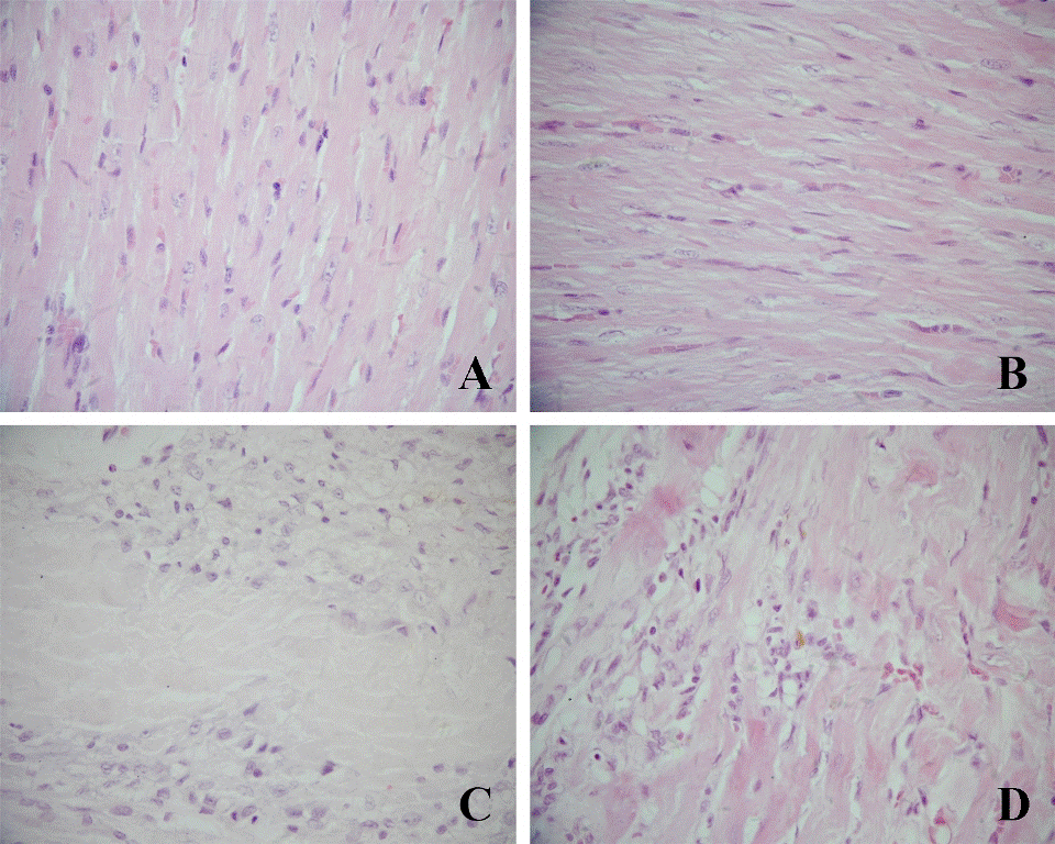
| Figure 1: The HE stained sections of myocardium in different groups. Myocardial cells in both sham rats (A) and sham-tumor rats (B) were lined in order with clear nuclear staining. Myocardial cells in both AMI-tumor rats (D) and AMI rats (C) were atrophied with necrosis and surrounded by the large number of new granulation tissue (400X). |