 |
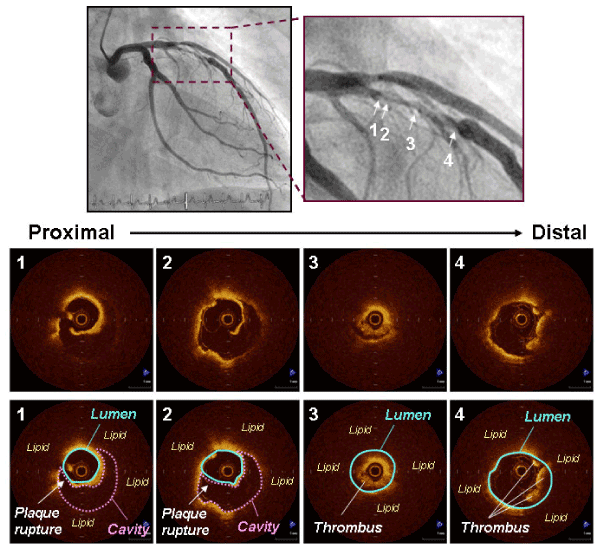
| Figure 1: Representative OCT images of plaque rupture in unstable angina pectoris. Angiography showed a complex lesion in the left anterior descending coronary artery. OCT revealed a plaque rupture with a residual fibrous-cap and plaque cavity (#1 and #2), and intracoronary thrombi (#3 and #4). Schemas demonstrate plaque rupture, lumen, ruptured cavity, lipid, and thrombi. Plaque rupture was observed at the minimal lumen area site [minimal lumen area = 1.83 mm2 (#1); maximal ruptured cavity area = 3.26 mm2 (#2)]. The plaque was considered to contain abundant lipid (number of lipid quadrants = 4). Thrombi occupied coronary lumen distal to the plaque rupture. |