|
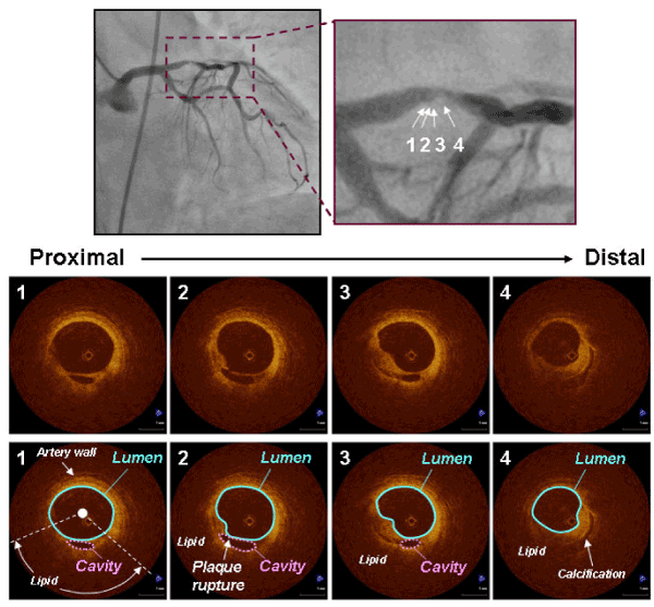
| Figure 2: Representative OCT images of plaque rupture in stable angina pectoris. Angiography showed an eccentric stenosis in the left anterior descending coronary artery. OCT revealed a plaque rupture with a residual fibrous-cap and plaque cavity (#1, #2, and #3). Schemas demonstrate plaque rupture, lumen, ruptured cavity, 3-layered coronary artery wall, lipid, and calcification. Plaque rupture was located proximally to the minimal lumen area site [maximal ruptured cavity area = 0.45 mm2; (#2) minimal lumen area = 2.59 mm2 (#4)]. The plaque contained lipdic tissue (number of lipid quadrants = 2) and calcium. Intracoronary thrombi were not detected in the lesion with plaque rupture. |