|
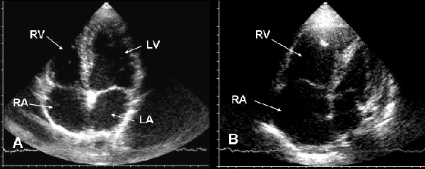
| Figure 1: Echocardiography. A: Apical 4-chambers scan of a normal heart. Right (RA) and left (LA) atrium show the same dimension. Left ventricle (LV) is near 2/3 larger than right ventricle (RV). B: Apical 4-chambers scan in a shocked patient. Right chambers are enlarged suggesting acute pulmonary embolism. |