 |
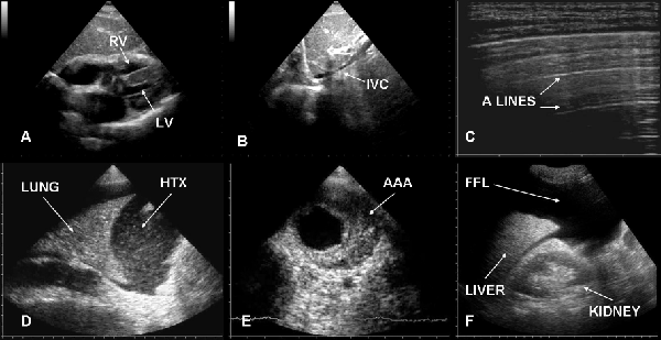
| Figure 9: Ultrasound in cardiac arrest does to hypovolemia. Typically cardiac chambers are small and hyperkinetic (A), inferior vena cava is slight (B) and lung is “dry” (C). Ultrasound can also detect some causes of hypovolemia such as large hemothorax (D), abdominal aortic aneurysm (E) or abdominal free fluid (F). RV=right ventricle, LV=left ventricle, IVC=inferior vena cava, HTX=hemothorax, AAA=abdominal aortic aneurysm, FFL=free fluid. |