|
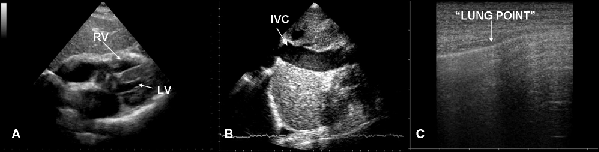
| Figure 12: Ultrasound in cardiac arrest does hypertensive pneumothorax. Typically cardiac chambers are small and hyperkinetic (A), inferior vena cava is dilated (B) and lung examination show the absence of sliding and B lines and in non massive pneumothorax the presence of the “lung point” reflecting the border between aerated lung and pneumothorax (C). |