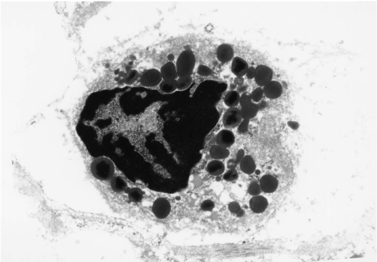
Figure 3:
Electron microscopic photograph of an eosinophil infiltrating the adventitia from our case No. 1. Characteristic dense granules are observed in the cytoplasm.