 |
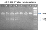
| Figure 1: The electrophoresis patterns of the AIF-1 genotypes after RE digest are shown. M indicates the molecular marker (50-bp). A 414 bp fragment was observed using generic primers flanking -932. Amplification and PML-1 digestion resulted in two fragments (227 base pairs and 187 base pairs) that confirm the C→T substitution. |