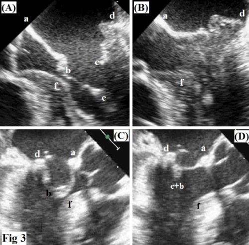
a. anterior annulus, b. tip of anterior leaflet, c. tip of posterior leaflet, d.
posterior annulus, e. papillary muscle, f. ventricular septum.
(A) Pre-mitral valve repair during systole. Anterior leaflet is redundant, the second order chords seem to be slack so the tip of the leaflet kisses the bulging septum.
(B) Imagine demonstrates the insufficiency of mitral valve due to a prolapsed and huge P2 of posterior leaflet.
(C) Post-mitral valve repair during diastole. During the end diastole, the paradoxical chords ensure the avoidance of contact between the tip of the anterior leaflet and the bulging septum. The annuloplasty ring seems to be downsized. The image shows a large
surface of coaptation between the two leaflets (c+b).