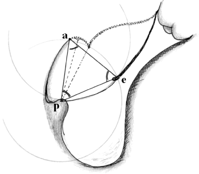
a. Posterior annulus (5 or 7 o’clock position), p. Papillary muscle, e. Edge
of anterior leaflet (5 or 7 o’clock position). The triangle a.p.e is an
equilateral triangle.
The plane of the posterior wall of left ventricle outflow tract extends from
the tips of the papillary muscles to the anterior annulus of mitral valve. In the
physiological condition this plane is curved slightly during the end diastole.
The length of the first order native chord of the anterior leaflet “p-e” has
approximately the same length of the first and second order of the posterior
leaflet. Therefore, the distance between the posterior annulus and the tip of
appropriate papillary muscle (not always the third order chord of posterior
leaflet) is equal to the length of the first order chord of the anterior leaflet
which is located at the same cross profile level. The tip of papillary muscle
“p” is considered the vertex of the angle between the posterior annulus–tip
of papillary muscle ray “p-a” and the first order chord of the anterior leaflet
ray “p-e” during the end diastole “ filling angle”, and by implanting a ring
annuloplasty this angle is narrowed. Thus, to obtain the filling angle wide
as much as possible, the triangle a.p.e during the end diastole should be
an equilateral triangle, so e-a (new chord) equal to e-p ( first order chord
of anterior leaflet).