|
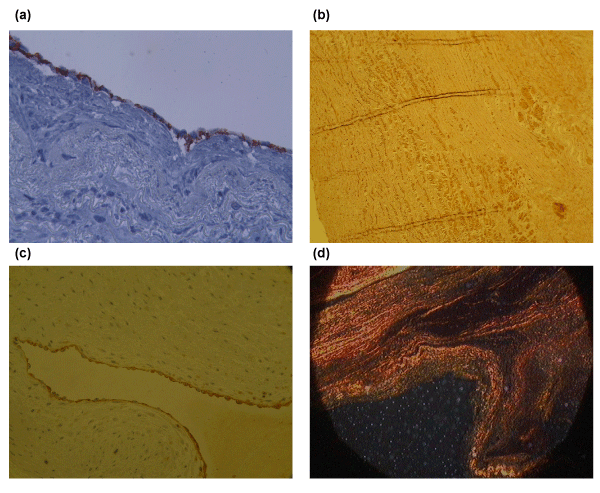
| Figure 2: A represantative sample of an implanted decellularized xenograft after 4 months of implantation. a) CD 31 staining demonstrates a confluent monolayer of endothelial cells at the wall of the decellularized xenograft. b) Factor VIII staining showed a monolayer of endothelial cells at the leaflets. c) Von Kossa shows a lack of calcification of the decellularized xenograft. d) Serius red staining demonstrates the remodeling potential of the collagen.. |