|
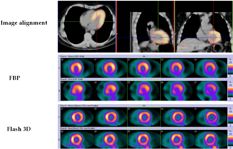
| Figure 1: The resulting image from FBP and Flash 3D reconstruction. Top row shows the alignment of cardiac data matching CT and SPECT images. Middle row shows midventricular short-axis slices reconstructed with FBP. Note the presence of an inferior wall perfusion defect on stress-rest images indicating an inferior wall myocardial infarction. Bottom row shows midventricular short axis slices reconstructed with Flash 3D. Note the absence of the inferior wall defect, suggesting that this was an artifact. Also, better contrast between the left ventricular walls and cavity and better visualization of the right ventricle. |