|
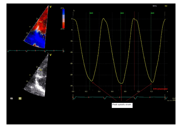
| Figure 4: Analysis of strain by tissue Doppler from the mid segment of the right free wall obtained from the apical 4-chamber view in a healthy term neonate. Upper left Figure: Strain by tissue Doppler image. Lower left Figure: Grey-scale image. Right Figure: Strain curve for three consecutive heart beats. Red arrows mark the peak systolic strain values. |