 |
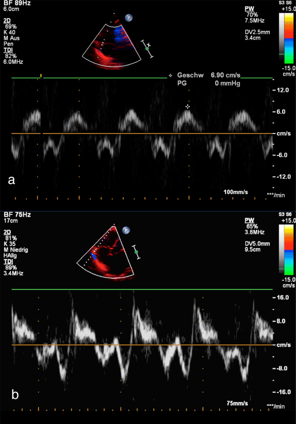
| Figure 6: Apical 4-chamber view. The white broken line indicate M-Mode cursor placement at the tricuspid lateral annulus. Representative image of the tricuspid annular peak systolic velocity (TAPSV) in a neonate (Figure 1a), and in a 15 year old adolescent (Figure 1b), respectively, with normal right and left ventricular function. |