|
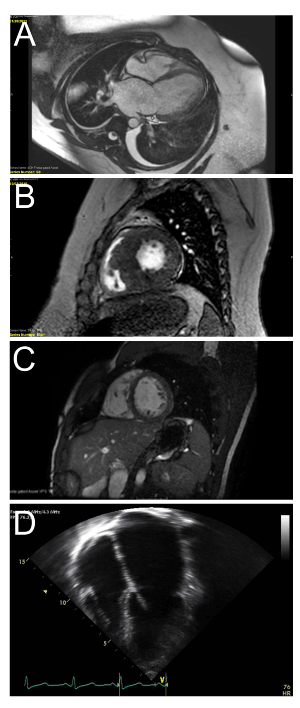
| Figure 5: A) Cardiac magentic resonance imaging of an adolescent with dilated cardiomyopathy depicting a dilated left ventricle in end-diastole with thinning of the left ventricular septum and free wall. B) Cardiac magnetic resonance imaging of an adolescent with hypertrophic cardiomyopathy depicting a thickened left ventricle with below normal left ventricular end diastolic dimension. C) Cardiac magnetic resonance imaging of a child with left ventricular noncompaction depicting trabeculations in the left ventricle with deep recesses. D) Echocardiogram of an adolescent with restrictive cardiomyopathy depicting severe dilation of both atria with preserved left ventricular size and thickness. |