|
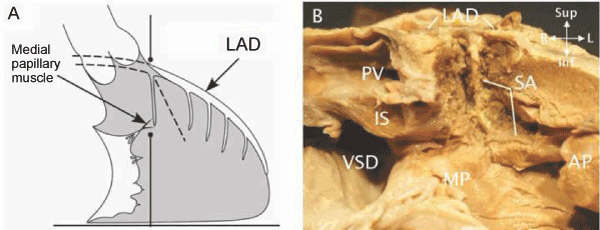
| Figure 5: A The 1st septal artery branches from the anterior descending artery (LAD) near the apical edge of the pulmonary root and passes through the anterior septum and septal band to reach the base of the medial papillary muscle (papillary muscle of the conus) (MP) According to Hosseinpour and colleagues [13], the position of the artery corresponds to a line constructed from the base of the MP perpendicular to the diaphragmatic wall. (Modified from Hosseinpour et al. [13] with permission). B A dissection of a heart with TOF showing the position of the 1st septal artery (SA). In this case the SA is deep in the anterior septum and septal band. It veers toward the apex and supplies the anterior papillary muscle (AP). In this case the line described in A would be basal to the artery. The artery is far from the VSD or any muscle likely to be resected during relief of outflow obstruction. |