 |
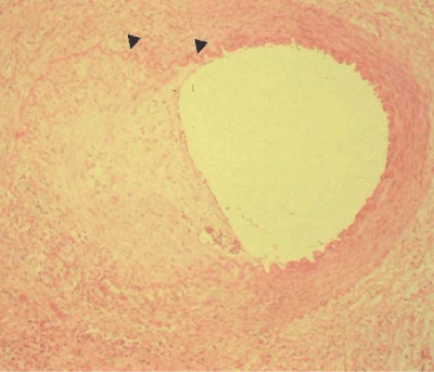
| Figure 25: A developing coronary artery aneurysm in a patient dying of Kawasaki disease. The portion of the vessel wall to the right is intact. On the left the vessel wall has been dissolved by lytic enzymes. The internal elastic lamina (arrow head) is disrupted. Disruption of the vessel wall allows the aneurysm to develop. Photomicrograph courtesy of Dr. Jane C. Burns, Director, Kawasaki Disease Research Center University of California San Diego. |