 |
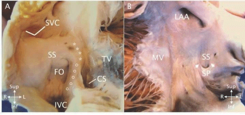
| Figure 1: Anatomy of the normal atrial septum. A - Opened right atrium showing the entrance of the superior vena cava (SVC), inferior vena cava (IVC), and coronary sinus (CS). The fossa ovalis (FO) forms the central part of the atrial septum and is bounded superiorly and rightward by septum secundum (SS). Septum primum is the thin floor of the fossa. The muscular base of the atrial septum (ο) is between the fossa and the coronary sinus. The AV canal septum (*) is adjacent to the tricuspid valve (TV). B - On the left atrial side, septum primum (SP) forms a hammock - shaped structure and has insertions (white arrows) on septum secundum (SS). LAA - left atrial appendage; MV - mitral valve. |