 |
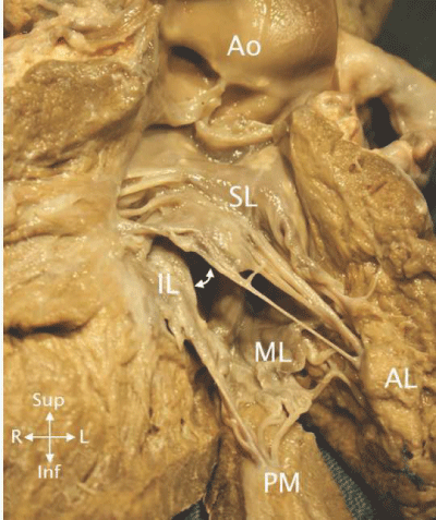
| Figure 5: The superior (SL) and inferior (IL) components of the anterior mitral leaflet as well as the mural leaflet (ML) are seen in the opened LV in this heart with ASD1. The superior leaflet attaches only to the anterolateral papillary muscle (AL) and the inferior leaflet attaches only to the posteromedial papillary muscle (PM). The mural leaflet attaches to both. The cleft (white double headed arrow) is the space between the superior and inferior leaflets. Only the superior leaflet is in continuity with the aortic valve (Ao). Chordal attachments of this leaflet high in the outflow can cause subaortic stenosis. |