 |
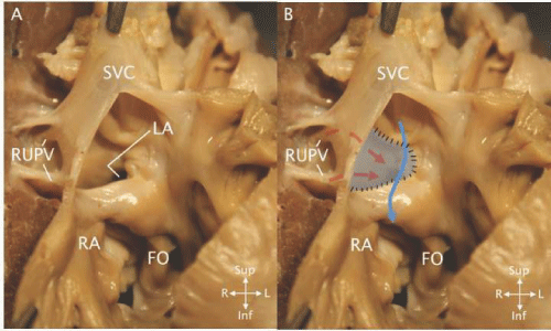
| Figure 6: Superior vena cava type of SVD. A - The right atrium (RA) is opened to show the junction with the superior vena cava (SVC). The left atrium (LA) can be seen through the SVD. Two right upper pulmonary veins (RUPV) drain to the SVC - RA junction near the defect. The fossa ovalis (FO) is seen inferior and leftward of the SVD. B - The same view showing a ‘patch’ (gray) covering the defect. Pulmonary venous blood (dashed red arrows) flows to the LA behind the patch while the blue solid arrow indicates SVC flow to the RA in front of the patch. |