 |
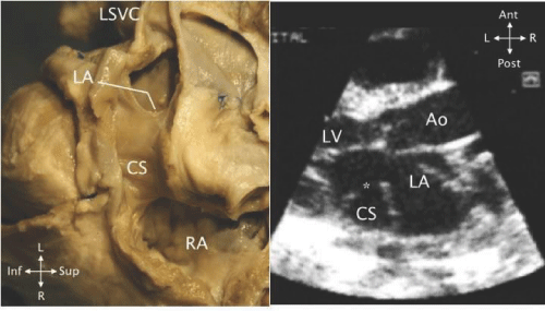
| Figure 7: Posterior view of a heart with a CSD. The coronary sinus (CS) has been opened from the left superior vena cava (LSVC) to the right atrium (RA). The left atrium (LA) can be seen through the CSD, an opening in the wall between the CS and the LA. B - An echocardiogram in parasternal long - axis view showing a CSD (*) between the dilated coronary sinus (CS) and the left atrium (LA). Ao - aorta; LV - left ventricle. |