|
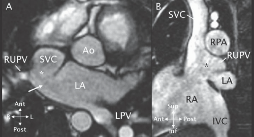
| Figure 10: A - A MRI 3D SSFP axial view showing a superior vena cava (SVC) type of SVD (*). The right upper pulmonary vein (RUPV) drains to the SVC - right atrial junction near the SVD. The orifice of the RUPV into the left atrium (LA) (white arrow) is the interatrial communication. B - Sagittal projection of the same data set shows the defect (*) between the SVC - RA junction and the RUPV. Ao - aorta; IVC - inferior vena cava; LPV - left pulmonary vein; RPA - right pulmonary artery. (Reprinted from Valente et al. [11] with permission). |