|
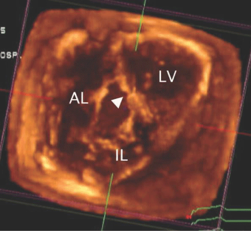
| Figure 15: A 3 - dimensional echocardiogram in a patient with Ebstein anomaly viewed from the apex. The septal tricuspid leaflet did not delaminate from the septum (arrowhead) leaving a gap between the anterior (AL) and inferior (IL) leaflets. LV - left ventricle. (Image courtesy of Jerry Marx, MD, Childrens Hospital Boston). |