|
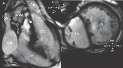
| Figure 19: MRI cine SSFP frontal (A) and short - axis (B) views in a patient with CTGA. A - The right - sided right atrium (RA) is aligned with the right - sided left ventricle (LV) and the LV is aligned with the right - sided main pulmonary artery (MPA). There is dephasing in the MPA (arrowhead) due to pulmonary stenosis. The aorta (Ao) is superior and leftward and aligned with the left - sided right ventricle (RV). B - The LV, marked by the smooth septal surface and free wall papillary muscles, is anterior while the coarsely trabeculated RV is posterior. SVC superior vena cava. |