|
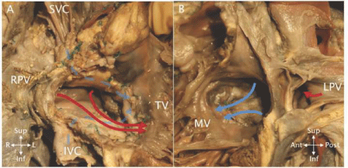
| Figure 21: A - The opened pulmonary venous atrium in a heart after a Mustard atrial switch operation. The red curved arrows indicate flow from the right (RPV) and left pulmonary veins toward the tricuspid valve (TV) and right ventricle. The dashed blue curved arrows indicate flow from the superior vena cava (SVC) and inferior vena cava (IVC) behind the limbs of the baffle toward the mitral valve and left ventricle. B - The opened systemic venous atrium in the same heart showing the other side of the baffle with SVC and IVC flow (blue curved arrows) toward the mitral valve (MV). The left pulmonary veins (LPV) are seen posterior to the systemic venous atrium. |