 |
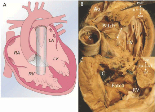
| Figure 23: A - a cartoon depicting the Rastelli operation for DTGA with ventricular septal defect (VSD) and pulmonary stenosis. The left ventricle (LV) is baffled to the aorta through the VSD and a conduit is placed from the right ventricle (RV) to the pulmonary artery. (Modified from Valente et al. [11] with permission). B - A heart specimen after a Rastelli operation shows the ventricular septal defect (double headed arrow) and the patch (Patch) directing the LV to the aorta (Ao). C – The opened RV showing the other side of the patch (Patch) and the junction of the RV. |