 |
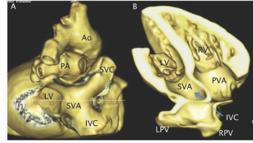
| Figure 25: 3D reconstruction of a CT exam of a waxed human heart with severe pulmonary venous pathway obstruction (*) after a Senning atrial switch operation. A - posterior view with the pulmonary veins removed to show the superior vena cava (SVC) and inferior vena cava (IVC) pathways joining the systemic venous atrium (SVA). The junction (*) between the portion of the pulmonary venous atrium that receives the right (RPV) and left (LPV) pulmonary veins and the supra - tricuspid portion of the pulmonary venous atrium (PVA) is severely stenosed. B - A cut in the plane indicated by the dashed white line in A showing the two portions of the pulmonary venous atrium. The IVC pathway (blue arrow) passes beneath the narrowed junction. |