|
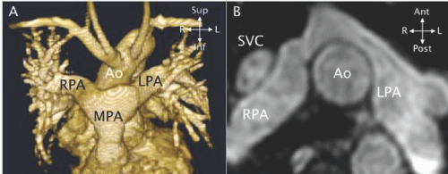
| Figure 26: CMR exam in two patients after an arterial switch operation. A - Frontal view of a 3 - D reconstruction from a MRA showing the anterior pulmonary artery (MPA) with the branch pulmonary arteries (RPA, LPA) on either side of the ascending aorta. (Reprinted from Valente et al. [11] with permission). B - A 3D SSFP in axial projection showing the distorted contours and proximal narrowing of the branch pulmonary arteries (RPA, LPA). The ascending aorta (Ao) is seen between the branch pulmonary arteries and the superior vena cava (SVC) in front of the RPA. |