 |
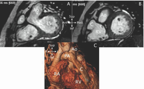
| Figure 27: CMR exam in a patient after a Rastelli operation. A - A cine SSFP short - axis cut through the ventricular septal defect (*) showing the flow path from the left ventricle (LV) to the aorta (Ao). B - A more apical cut showing the junction of the conduit (C) with the right ventricle (RV). C - A 3 - D reconstruction from the MRA showing the RV - pulmonary artery (PA) conduit (C) to the right of the Ao. The conduit is flattened by the chest wall. |