 |
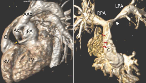
| Figure 28: 3D reconstructions from a MRA performed in an adult patient with complex double outlet right ventricle who underwent repair in childhood. A - Left lateral view showing a Damus - Kaye - Stansel anastomosis (*) between the pulmonary root (PA) and the ascending aorta (AAo) because of severe subaortic stenosis (yellow arrow). The left ventricle (LV) was baffled to the pulmonary root (PA) through the ventricular septal defect. B - A conduit (C) was placed between the right ventricle (RV) and the pulmonary arteries. There is severe proximal left pulmonary artery (LPA) stenosis (white arrowhead). The irregularities in the anterior wall of the conduit (red arrows) are artifacts from sternal wires. RPA - right pulmonary artery. |