 |
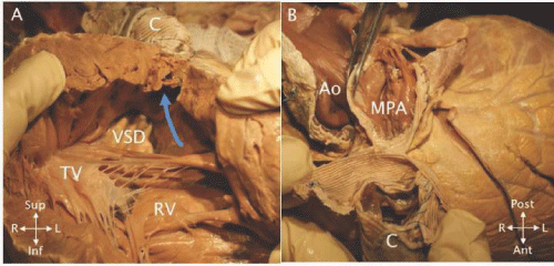
| Figure 30: A - Opened right ventricle (RV) in a patient with TOF and pulmonary atresia who underwent repair using a valved conduit. The RV outflow (blue curved arrow) was created from the ventriculotomy to which the conduit (C) was sewn. The ventricular septal defect patch (VSD) is seen extending above the tricuspid valve (TV). B - An anterior superior view of the same heart showing the connection of the valved conduit (C) to the main pulmonary artery (MPA) to the left of the aorta (Ao). |