|
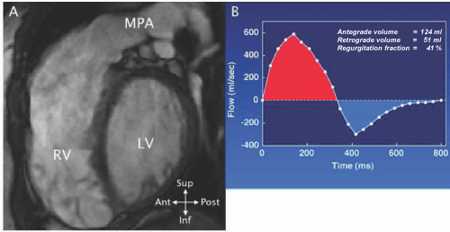
| Figure 33: A - MRI cine SSFP short - axis image in a patient late after repair of TOF. The RV is dilated due to severe pulmonary insufficiency from wide patch plasty of the RV outflow. B - Graph of instantaneous flow velocity vs time calculated from a phase contrast MR sequence prescribed across the right ventricular outflow. Antegrade flow is shown above baseline (red) and retrograde or regurgitant flow below (blue). The area under each portion of the curve indicates volume flow which is used to calculate the regurgitant fraction. |