 |
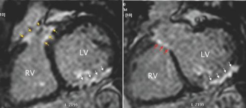
| Figure 34: MRI short - axis images showing delayed gadolinium enhancement in a patient after repair of TOF. A - A cut at the level of the right ventricular (RV) outflow showing delayed enhancement of the outflow patch (yellow arrows). The patient suffered a perioperative inferior infarction of the left ventricle (LV) indicated by delayed enhancement (white arrows). B - A cut through the LV outflow showing delayed enhancement of the patch closing the ventricular septal defect (red arrows). The LV inferior infarction is seen in this cut as well (white arrows). |