 |
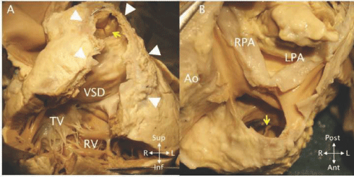
| Figure 36: A - The opened right ventricle (RV) in a heart following repair of TOF and implantation of a bioprosthesis (yellow arrow) in the pulmonary position. The ventricular septal defect patch (VSD) is completely endothelialized. The RV anterior wall (white arrowheads) is thin and fibrotic. This tissue is often removed when RV remodeling is performed during valve implantation. B - The bifurcation of the pulmonary artery showing the branches (RPA, LPA) and the arterial surface of the bioprosthesis (yellow arrow). |