 |
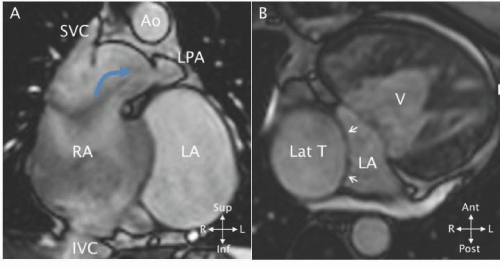
| Figure 40: A - MRI cine SSFP coronal view in a patient with an atriopulmonary connection (blue curved arrow). The right atrium (RA) is dilated and the atrial septum bulges into the left atrium (LA). B - MRI 3D SSFP axial view in a patient with functionally one ventricle (V) after a lateral tunnel Fontan operation. The lateral tunnel (Lat T) is seen in cross - section within the atrium. A patch (white arrows) separates the tunnel from the remainder of the atrium (LA). IVC - inferior vena cava; LPA - left pulmonary artery; SVC - superior vena cava. (Reprinted from Valente et al. [11] with permission). |