|
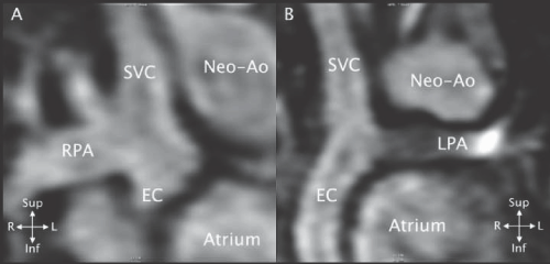
| Figure 41: MRI 3D SSFP frontal views in a patient with hypoplastic left heart syndrome after extracardiac conduit Fontan operation. A - The Glenn anastomosis between the superior vena cava (SVC) and the right pulmonary artery (RPA) is seen. The junction of the extracardiac conduit (EC) with the inferior aspect of the RPA is seen but the rest of the conduit is out of the plane. The neo - aorta (Neo - Ao) is to the left of the SVC. B - The conduit (EC), SVC and left pulmonary artery (LPA) are seen but the RPA is out of plane. As frequently seen the LPA is smaller than the RPA. Note that the conduit is separate from the atrium. |