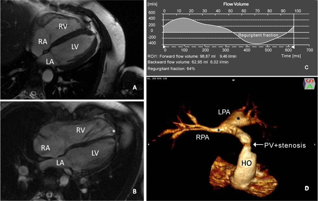
A: 4 chamber view cine imaging with normal size of RV and LV.
B: Enlarged RV and normal sized LV in a patient with pulmonary regurgitation after repair of TOF with transanular patch plasty. The apex is formed by the RV (*) while in a normally sized heart, it is formed by the LV.
C: Volume change over time from flow measurement of the pulmonary trunk showing severe pulmonary regurgitation with 64% regurgitant fraction.
D: VR 3D Reconstruction of the right ventricle and pulmonary arteries from MR angiography. Valvular and supravalvular stenosis in the homograft (HO) and stenosis of the RPA (+) with aneurysmatic enlargement of the LPA (*) in a patient with TOF after corrective surgery with pulmonary valve replacement by homograft.