|
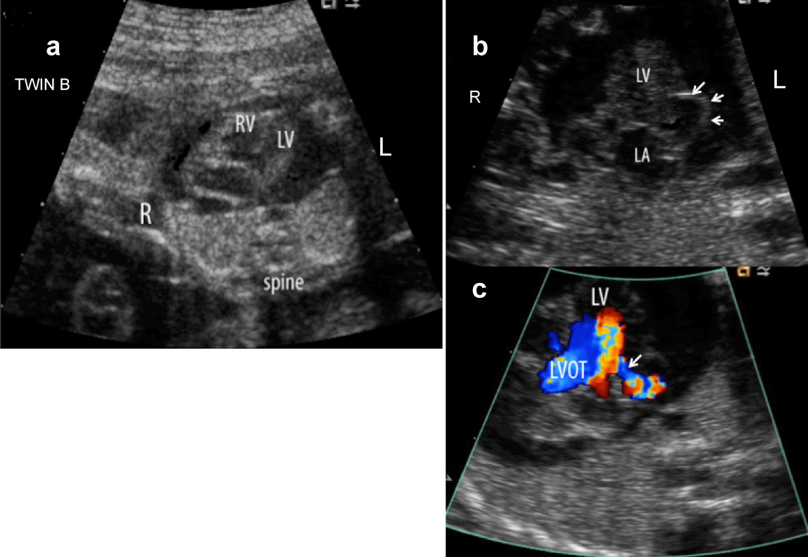
| Figure 3: LV free wall aneurysm versus diverticulum in a 20 week twin fetus. a) A massive pericardial effusion prompted initial referral for fetal echocardiography. b) On further imaging, a thin outpouching was demonstrated (arrows) arising from the lateral wall of the left ventricle (LV) which communicated through a small neck with the LV cavity. c) During ventricular systole, blood entered this hypokinetic aneurysm (arrow at neck). Spontaneous rupture led to an acute demise of this fetus at 24 weeks. L-left, R-right, LV-left ventricle, LA-left atrium, RV-right ventricle, LVOT-left ventricular outflow tract. |