 |
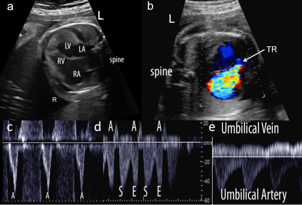
| Figure 4: 28 week gestational age fetus with evolving hydrops secondary to noncompaction cardiomyopathy and an Ebsteinoid tricuspid valve with severe tricuspid regurgitation. a) Significant trabeculations of the left ventricle are apparent from the 4 chamber view as are pleural and pericardial effusions. b) By color Doppler, tricuspid regurgitation (TR) is seen originating from low in the right ventricular cavity. c) Ventricular inflow was of short duration and nearly uniphasic suggesting the presence of diastolic pathology. d) Significant flow reversal in atrial systole in the ductus venosus and e) umbilical venous pulsations were present and in keeping with increased central venous pressures. RA-right atrium, LA-left atrium, LV-left ventricle, RV-right ventricle, L-left, R-right, TR-tricuspid regurgitation, E-early diastolic flow, S-flow in ventricular systole, A-flow in atrial systole |