|
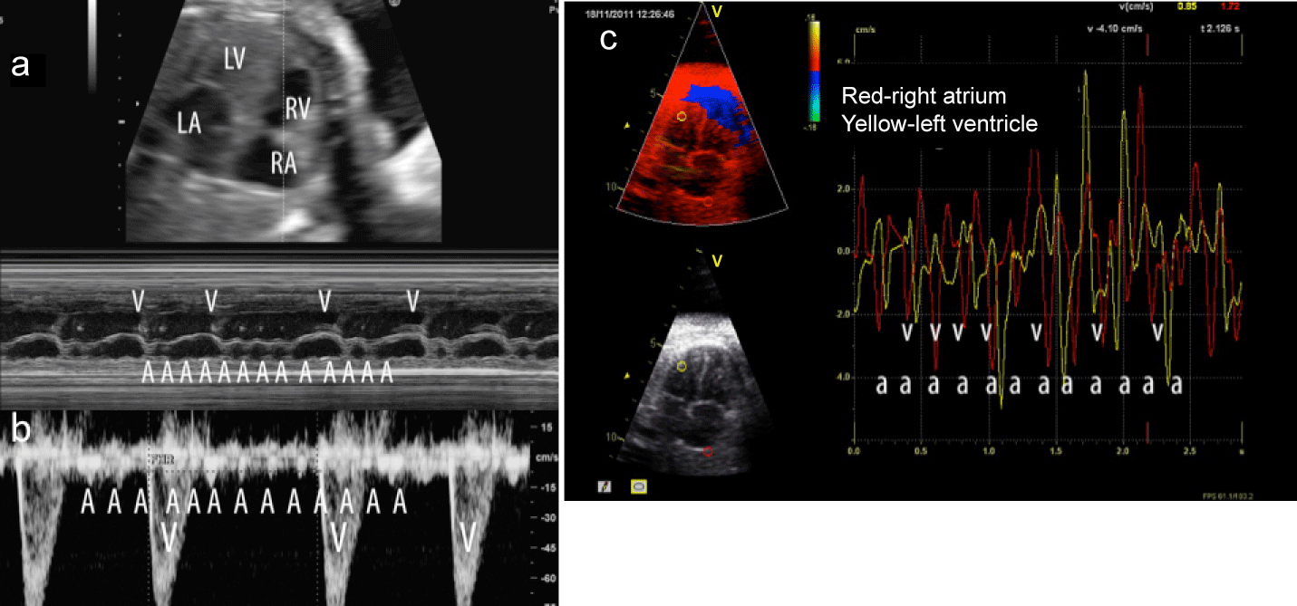
| Figure 5: Assessment of a 27 week fetus with atrial flutter and variable A-V conduction. a) M mode tracings demonstrate the timing of atrial (A) and ventricular (V) contractions. b) Simultaneous superior vena cava and ascending aortic pulsed Doppler spectra show timing of a wave reversal in atrial systole in the superior vena cava (A) and forward flow in the aorta during ventricular systole (V). c) Tissue color Doppler tracings also demonstrate the temporal relationship between atrial (A) and ventricular (V) contractions by changes in myocardial velocities. The atrial rate(A-atrial contraction or systolic flow) is roughly 300bpm whereas the ventricular rates (V-ventricular contraction or systolic flow) range from 100-200bpm. A-atrial systolic contraction or flow, V-ventricular contraction or forward aortic flow in ventricular systole. Maternal sotalol therapy successfully treated this rhythm with subsequent vaginal delivery at 40 weeks. LA-left atrium, LV-left ventricle, RA-right atrium, RVright ventricle |