 |
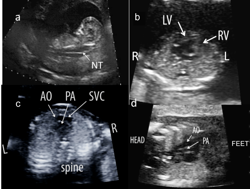
| Figure 6: 12 and 3/7 weeks gestational age fetus with a) a 7mm nuchal translucency (NT). b) On further assessment, the four chamber revealed levocardia inverted ventricles and a mildly foreshortened left sided right ventricle. At 14 weeks, the presence of L-transposition (“corrected transposition”) of the great arteries demonstrated by c) an abnormal 3 vessel view with an anterior and leftward aorta arising from the right ventricle and posterior pulmonary artery from the left ventricle and d) parallel arrangement of the great arteries. L-left, R-right, AO-ascending aorta, PA-pulmonary artery, LV-left ventricle, RVright ventricle, SVC-superior vena cava |