|
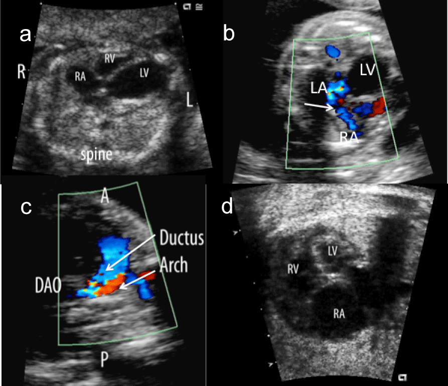
| Figure 8: Images from a 21 week fetus with critical aortic stenosis. a) At this stage in addition to a thickened and hypoplastic aortic valve, the left ventricle (LV) was found to be severely dilated and akinetic. There was b) left to right atrial flow and c) retrograde aortic arch flow in keeping with high left heart filling pressures and severely reduced left ventricular output, respectively. With redistribution of blood flow to the right heart and decreased filling of the left ventricle, there was progressive left heart hypoplasia such that by 35 weeks the fetus had findings consistent with hypoplastic left heart syndrome. LA-left atrium, RV-right ventricle, RA-right atrium, L-left, R-right, DAo-descending aorta. |