|
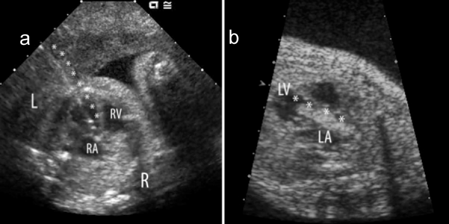
| Figure 9: Fetal catheter intervention for severe aortic valve stenosis at 25 weeks. a) After positioning of the fetus to ensure the left ventricular apex was oriented towards the maternal abdominal wall, a needle was used to puncture through the maternal abdomen, uterus, fetal chest and fetal left ventricular apex (*course of the needle). A wire was then passed through the needle into the left ventricular outflow tract and through the aortic valve. c) A coronary balloon catheter was subsequently guided over the wire through the aortic valve and expanded to dilate the valve (*course of balloon). LA-left atrium, RAright atrium, LV-left ventricle, RV-right ventricle, L-left, R-right. |