|
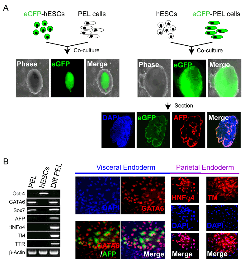
| Figure 2: Confirming the PEL identity. (A) The PEL cells constitutively emerged from their clonally-related hESCs maintained under the defined culture conditions segregate to appropriate extra-embryonic layer location when co-culturing with undifferentiated hESCs. Whether one created the chimeric aggregates by mixing green-fluorescent PEL cells (eGFP+) with white-non-fluorescent hESCs or white-non-fluorescent PEL cells with greenfluorescent hESCs (eGFP+) in suspension culture, the PEL cells established residence at the periphery of the chimeric and expressed endoderm-specific markers, e.g., AFP (red). DAPI is blue. (B) RT-PCR analysis of early PE associated markers (GATA4/6 and SOX7) and late markers associated with differentiation into visceral (alpha-feto protein [AFP], transthyretin [TTR], HNFα4) and parietal (HNFα4, thrombomodulin [TM]) endoderm in PEL cells (PEL), undifferentiated hESCs (hESCs), and differentiated PEL cells treated with BMP2 and ddcAMP (Diff PEL). Immunocytochemical analysis of hESCderived PEL cells differentiating into cells expressing visceral or parietal endoderm markers. Prior to differentiation, all the PEL cells were GATA6+ (red), but AFP-negative. Following treatment with BMP2 and ddcAMP, ~90% of cells become positive for HNFα4 (red) and TM (red) and ~10% of cells become positive for AFP (green). Note that the AFP+ cells (green) began to emerge from the GATA6+ (red) PEL cells such that the 2 markers now colocalized within the same cells. |