|
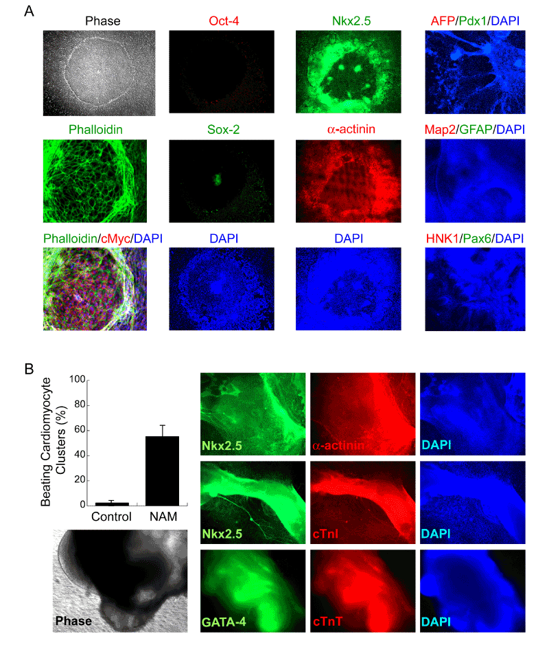
| Figure 5: Direct induction of clinically-suitable human myocardial grafts from biologics-free hESCs. (A) Upon exposure of undifferentiated biologics-free hESCs to NAM under the defined culture system, large differentiated phalloidin (green)-positive, cMyc (red)-positive, Oct-4 (red)- negative, and Sox-2 (green)-negative cells within the colony began to emerge. NAM-induced Oct-4-negative (red) cells began to express Csx/Nkx2.5 (green) and a-actinin (red), consistent with early cardiac differentiation. Progressively increased intensity of Nkx2.5 was usually observed in areas of the colony where cells began to pile up. These differentiated cells did not express markers for other lineages, including AFP (red), Pdx1 (green), Map-2 (red), GFAP (green), HNK1 (red), and Pax6 (green). All cells are indicated by DAPI staining of their nuclei (blue). (B) These biologics-free hESC-derived cardioblasts progress to beating cardiomyocytes with high efficiency. NAM-induced cardiac-committed biologics-free hESCs yielded beating cardiomyocytes with high efficiency, as assessed by the percentages of cellular clusters that displayed rhythmic contractions. Quantitative data are mean values from at least three separate experiments and at least 2 different cell lines. Germ-layer-induced hESCs without treatment were used as controls. The representative large beating cardiomyocyte clusters (phase) were immunopositive for Nkx2.5 (green), GATA-4 (green), a-actinin (red), cardiac troponin I (cTnI, red), cardiac troponin T (cTnT, red) (DAPI is blue). |