|
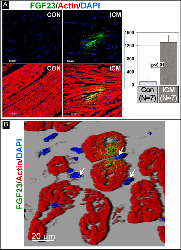
| Figure 3: FGF23 in cardiomyocytes of patients with ischemic cardiomyopathy. (A) Confocal images and morphometrical analysis of FGF23 in human myocardial tissue of healthy controls (CON) and of patients with ischemic cardiomyopathy (ICM). (B) FGF23 positive cardiomyocytes at a higher magnification. Note the strong staining ofcardiomyocytes and the weak signals of FGF23 in the nuclei of non-cardiomyocytes. |