 |
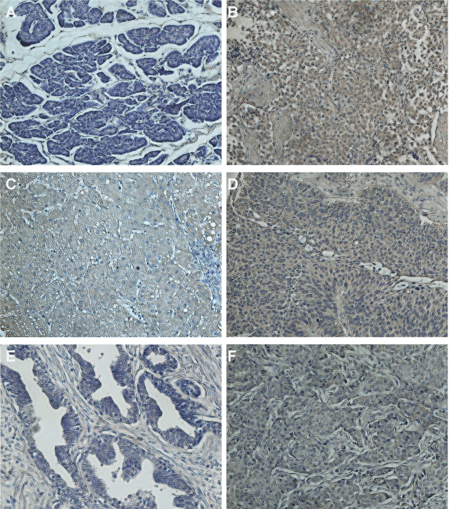
| Figure 5: Immunohistochemical analysis of expression of EDDR1 in normal and cancer tissues: Tissue arrays comprising of matched normal or cancer specimens (breast, ovarian and prostate tissues) and a panel of normal tissues (Figure S1: duodenum mucosa, spleen, pancreas, liver, lymph node and uterus myometrium) were stained with a EDDR1 specific monoclonal antibody followed by treatment with an HRP-conjugated secondary antibody reagent. Slides were developed with DAB substrate and tissues were counterstained with Meyer’s hematoxylin. Stained tissue sections were analyzed using a fluorescent microscope and micrographs were captured at 200× magnification. |