 |
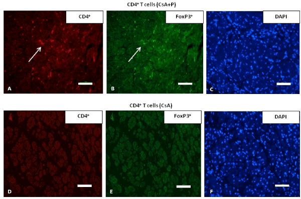
| Figure 6: Immunoflourescent staining for regulatory cells (Tregs) in animals conditioned with CD4+ T cells. (A, B, C) Cardiac allograft tissue stained with anti-CD4+ (red), anti-Foxp3+ (green) antibody and DAPI, respectively, from animals that had received CsA+P - conditioned CD4+ T cells. Arrows denote cluster of CD4+ and Foxp3+ cell infiltration into the graft. (D, E, F) Cardiac allograft tissue from animals treated with CsA alone -conditioned CD4+ T cells, stained with anti-CD4 (red), anti-Foxp3 (green) antibody and DAPI, respectively. There is no infiltration with CD4+ and Foxp3+ positive cells. Bar is equal to 50μm. |