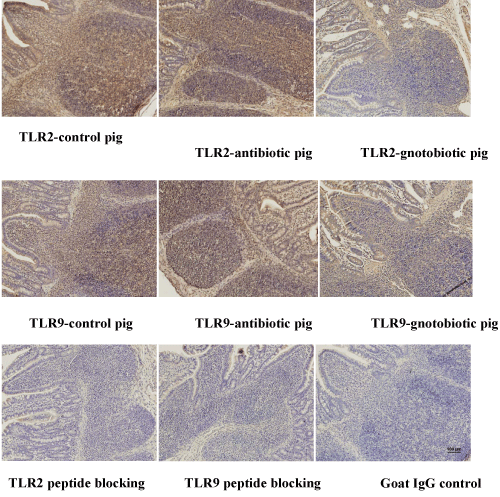
 Ileal tissue sections were prepared
and stained as described in the materials and methods. Briefly, tissue
sections were treated with Dako target retrieval solution for unmasking the
tissue antigens. After blocking, tissue sections were incubated with goat antihuman
TLR-2, -4, -5 and -9 antibodies. Normal goat IgG treatment served as
an isotype antibody negative control. Folllowing 3X washings, tissue sections
were incubated with HRP-conjugated rabbit anti-goat IgG antibody, treated
with DAB and counter-stained with Mayer’s hematoxylin. The sections were
dehydrated and mounted permanently. The specificity of each TLR antibody
was tested by pre-incubating TLR antibodies with the corresponding peptides
and this treatment completely abrogated the TLR staining. The tissue sections
from various treatment groups showed TLR-2 and TLR-9 specific staining. No
TLR-4 and -5 specific tissue staining was observed.
Ileal tissue sections were prepared
and stained as described in the materials and methods. Briefly, tissue
sections were treated with Dako target retrieval solution for unmasking the
tissue antigens. After blocking, tissue sections were incubated with goat antihuman
TLR-2, -4, -5 and -9 antibodies. Normal goat IgG treatment served as
an isotype antibody negative control. Folllowing 3X washings, tissue sections
were incubated with HRP-conjugated rabbit anti-goat IgG antibody, treated
with DAB and counter-stained with Mayer’s hematoxylin. The sections were
dehydrated and mounted permanently. The specificity of each TLR antibody
was tested by pre-incubating TLR antibodies with the corresponding peptides
and this treatment completely abrogated the TLR staining. The tissue sections
from various treatment groups showed TLR-2 and TLR-9 specific staining. No
TLR-4 and -5 specific tissue staining was observed. |