|
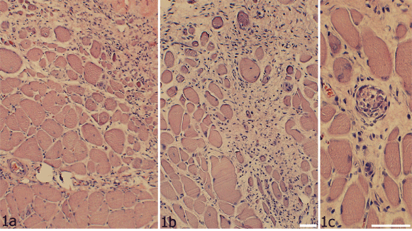
| Figure 1: Sections of specimens stained with H&E. The specimens were from the experimental (a) and non-experimental (contralateral) (b,c) sides of the 6w group. It is shown that pronounced morphological changes had occurred. Marked presence of loose connective tissue and presence of inflammatory infiltrates (above, a, and above and to the right, b) and occurrence of a variability in muscle fiber sizes are thus seen. Presence of a morphologically abnormal muscle fiber (necrotic muscle fiber), being completely infiltrated by cells, is shown in (c) (middle part). Bars= 25 μm. |